Acute Phase Reactants (ESR, CRP)
Last updated: November 25, 2014
Synonyms: ESR (Erythrocyte Sedimentation Rate), Sed Rate, CRP (C-Reactive Protein), hsCRP
CPT Codes: ESR 85652; CRP 86140; hsCRP 86141
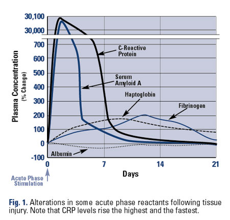
Description: Acute-phase reactants are a group of plasma proteins normally produced by the liver as part of an inate and adaptive immune response, driven by proinflammatory cytokines, namely interleutkin-1 (IL-1), IL-6 and tumor necrosis factor. Synthesis of these diverse proteins increases greatly in response to inflammatory stimuli in both acute and chronic settings. Examples of acute-phase reactants include (a) coagulation proteins (fibrinogen, prothrombin), (b) transport proteins (haptoglobin, transferrin, ceruloplasmin), (c) complement components (C3, C4), and (d) miscellaneous proteins [fibronectin, serum amyloid A (SAA), C-reactive protein (CRP), ferritin] (Figure 1). Acute-phase reactants are commonly used as an indirect measure of the extent of inflammation. For rheumatic diseases, the most commonly used acute-phase reactants are the CRP and the erythrocyte sedimentation rate (ESR). The ESR measures the rate of gravitational settling of erythrocytes and rouleaux formation, which is accelerated by a variety of factors, the most important of which is fibrinogen that lessens the negative charge between red blood cells (RBCs).
Method: In practice, the two most often used clinical tests for the acute-phase reactants are the ESR and CRP. The CRP is most often (and accurately) measured by rate nephelometry (an automated antigen/antibody-mediated reaction). Assays used for “highly-sensitive” CRP (hsCRP) tests are the same as conventional assays with the only difference being the lowest detectable levels. hsCRP is able to accurately measure CRP levels below 1 mg/ml. The ESR is performed by the Westergren method (recommended by the International Committee for Standardization in Hematology) and uses anticoagulated venous blood, diluted 4:1 with sodium citrate and placed in a 200-mm glass tube with a 2.5-mm internal diameter. At the end of 1 hour, the distance (in millimeters) from the meniscus (plasma and sodium citrate) to the top of the column of erythrocytes is recorded as the ESR. The modified Westergren method substitutes ethylenediaminetetraacetic acid (EDTA) for sodium citrate and enables the same sample of blood to be used for other tests. Both methods yield identical results.
Normal Values: CRP is normally <0.8 mg/dL (or < 8 mg/L) and is less affected by age or gender than the ESR. hsCRP is normally < 0.5 mg/dL (or 5.0 mg/L). It is critical to note and understand the units by which CRP is reported. Hence 10 mg/L is equal to 1.o mg/dL. Likewise a CRP of 2.0 mg/dL is elevated while 2.0 mg/L is not. In young men, the ESR ranges from 0 to 13 mm/h and from 0 to 20 mm/h in women. Importantly, the ESR increases with advancing age. Rough formulas for age-adjusted estimates of ESR are as follows: men: ESR = age in years/2; women: ESR = (age in years + 10)/2.
Increased in: CRP value rise faster than ESR. After a stimulus, CRP levels may rise up to 10,000 fold in several hours, peak in 48 hours, has a plasma half-life 19 hours, and shows no diurnal variation. The ESR rises and falls more slowly and is preferred by some in monitoring chronic inflammatory states. The ESR and CRP are increased in a variety of disorders including:
- Acute or chronic inflammatory disorders: Gout, rheumatoid arthritis (RA), rheumatic fever, spondyloarthropathies, polymyalgia rheumatica (PMR), giant cell arteritis (GCA) and other forms of vasculitis, inflammatory bowel disease, etc.
- Tissue injury/necrosis: Acute myocardial infarction, tissue ischemia or infarction, transplant rejection, malignant tumors, after surgery, burns, and trauma.
- Infections: Bacterial (e.g., endocarditis, osteomyelitis, intraabdominal infections) and some viral infections (e.g., acute viral hepatitis).
- Miscellaneous: May also be elevated in late pregnancy and postpartum, hyper- and hypothyroidism, azotemia and nephrotic syndrome. CPR levels can be increased by oral contraceptives, hormone replacement therapy, periodontal disease and smoking.
Not all patients with inflammatory or infectious disorders will manifest an elevated ESR or CRP. Between 35-45% of active RA, 20-30% of spondyloarthritis and 30-40% of active lupus patients will have normal ESR and CRP values. Many patients will show discordance with elevation of one (e.g., ESR), but not the other (e.g., CRP) – this is commonly seen in RA, infection, renal disease and patients with low albumin levels. Extreme elevations of the ESR and CRP are often seen in PMR, GCA, vasculitis, acute infections, Still’s disease, macrophage-activation syndrome and acute gout. Note: More than 10% of very high ESR (>75 mm/h) values lack an identifiable cause. This is particularly true in the elderly. The physician should wait and repeat the test in 3 to 6 months and not routinely embark on an exhaustive, expensive, or invasive search for an occult neoplastic, infectious, or inflammatory process.
Using hsCRP, patients with CRP in the upper one-third of normal range have a 2 fold risk of a future coronary event compared to those in the bottom third. The import of CRP in coronary artery disease is unclear. Atheromas and plaque instability have been associated with local inflammation. At the same time systemic inflammation may contribute to vascular disease (eg, increased rates of cardiovascular deaths in RA patients). Hence CRP may be a marker for a proatherogenic state and those with elevated levels may be predisposed to atherothrombotic events.
Decreased in: The ESR may be low with RBC abnormalities (polycythemia, microcytosis, spherocytosis, sickle cell, other hemoglobinopathies), hypofibrinogenemia, congestive heart failure, and cachexia and in those on high-dose corticosteroids or tumor necrosis factor inhibitors. CRP may be decreased with decreased by statin use, weight loss, physical exercise, and moderate alcohol use. CRP elevation is impaired with liver failure, but is unaffected by other comorbidities or organ dysfunction. The ESR and CRP may be normal in patients with systemic lupus erythematosus (SLE), polymyositis, scleroderma, pregnancy, osteoarthritis, and most viral infections. It is the goal of effective management to normalize or decrease acute phase reactant (ESR, CRP, etc.) measures in the course of antiinflammatory treatment.
Confounding Factors: The ESR may be increased by macrocytosis, hypercholesterolemia, increased fibrinogen, and high ambient temperatures. The ESR may be decreased by disorders of RBC morphology (see above), high leukocyte counts, hyperviscosity states, or a delay (>2 hours) in testing.
Indications: The ESR and CRP should not be used to screen asymptomatic individuals. They should be considered in the evaluation of febrile states, fatigue, weight loss or musculoskeletal complaints. The acute-phase reactants may be used in to distinguish inflammatory from noninflammatory disorders. Although nonspecific, they may be diagnostically or therapeutically useful. The American College of Physicians recommends that the ESR be used in the diagnosis and monitoring of polymyalgia rheumatica, temporal arteritis, and Hodgkin disease. With inflammatory disorders such as RA, the ESR should primarily be used to resolve conflicting clinical data (e.g., the patient whose examination improves but subjective complaints do not). Nonetheless, there is a long history of experience with the ESR in diagnosis and monitoring of inflammatory disorders such as RA. The CRP is useful in monitoring disease activity and response to therapy in chronic inflammatory disorders (e.g., RA). Some prefer the CRP to the ESR in monitoring inflammatory disease because changes are more acute. The CRP increases rapidly after an inflammatory stimulus and returns to normal within days; the ESR may take days to increase and may return to normal values over weeks. Also, the CRP is more dynamic (the difference between the levels seen in inflammatory and normal states is greater). Surgeons sometimes prefer to monitor the CRP and use very high elevations in CRP to indicate postoperative infection because the ESR may be elevated owing to the surgery itself. In inflammatory conditions associated with protein-losing nephropathy, such as SLE, the ESR may always be elevated, and the CRP may be a better indicator of superimposed infection.
Cost: ESR, $19–36; CRP, $25–55; ferritin, $45–65.
| Table 1: Comparison of CRP and ESR | ||||
| Acute-Phase Reactant | Advantages | Disadvantages | ||
|---|---|---|---|---|
| CRP | Rises and falls quickly; less age dependent | Results may take longer – 60 min to perform + lab time | ||
| Few confounding factors | Extensively studied in RA; less with other diseases | |||
| Uninfluenced by RBC shape | May be normal in the face of active inflammation or infection | |||
| Fewer technical errors | Slightly more expensive than ESR | |||
| ESR | Simple to perform; widely available | Rises and falls slowly | ||
| Greater use and familiarity | Age and gender dependent | |||
| Inexpensive | Many potential confounding factors | |||
| Done on-site in 60 minutes | Influenced by RBC morphology | |||
| CRP, C-reactive protein; RA, rheumatoid arthritis; RBC, red blood cell; SLE, systemic lupus erythematosus; ESR, erythrocyte sedimentation rate. | ||||
Comments: Although the CRP generally parallels the ESR, it increases earlier than the ESR (4–6 hours) and returns to normal first. It is especially useful in situations in which the ESR results may be affected by confounding variables (anemia, polycythemia, abnormal RBC morphology, hypergammaglobulinemia, and congestive heart failure). The CRP is superior to the ESR in assessing disease activity and therapy in RA (Table 1). The ESR is never diagnostic of a single disease, and trends may be more valuable than a single result. Extreme elevations (>100 mm/h) may suggest cancer, vasculitis (e.g., giant cell arteritis), adult-onset Still disease, spondyloarthropathy, or serious infections. Normal values do not exclude disease. It is not useful as a screening test in asymptomatic individuals.
BIBLIOGRAPHY
Sox HC, Liang MW. The erythrocyte sedimentation rate: guidelines for rational use. Ann Intern Med 1986;104:515–523. PMID:3954279
Otterness IG. The value of C-reactive protein measurement in rheumatoid arthritis. Semin Arthritis Rheum 1994;24:91–104. PMID:7839158
Pepys MB, Hirschfield GM. C-reactive protein: a critical update. Clin Invest 2003;111:1805–1812.PMID: 12813013
Costenbader KH, Chibnik LB, Schur PH. Discordance between erythrocyte sedimentation rate and C-reactive protein measurements: clinical significance. Clin Exp Rheumatol 2007; 25:746-749. PMID: 18078625
Berbari E, Mabry T, Tsaras G, Spangehl M, Erwin PJ, Murad MH, Steckelberg J, Osmon D. Inflammatory blood laboratory levels as markers of prosthetic joint infection: a systematic reviewand meta-analysis. J Bone Joint Surg Am. 2010;92:2102-9. PMID: 20810860
Vercoutere W, Thevissen K, Bombardier C, Landewé RB. Diagnostic and predictive value of acute-phase reactants in adult undifferentiated peripheral inflammatory arthritis: a systematic review J Rheumatol Suppl. 2011;87:15-9. PMID:21364051


