Arthrocentesis & Injections: Knee (Patellofemoral)
Last updated: October 15, 2014
Patient Position: Patient should lie supine and be made comfortable with the head supported and slightly inclined. If a flexion contracture exists, use pillows to support and position the knee so that the quadriceps is relaxed during the procedure.
Limb Position: The leg should be parallel to the ground and the knee may be slightly flexed. Remove the shoe and position the foot perpendicular to the ground so that the leg is neither externally nor internally rotated.
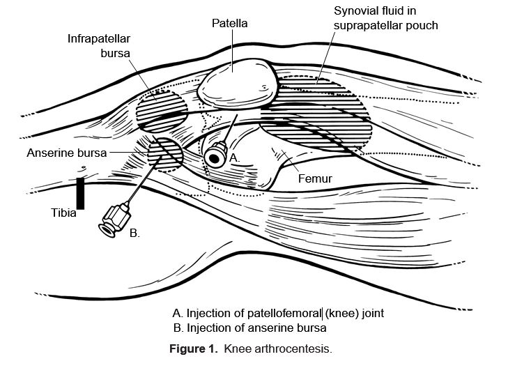
Bony Landmarks: Mark the medial, lateral, superior, and inferior borders of the patella (Fig. 1).
Site/Angle of Entry: Use a medial or lateral approach. Site selection should be based on maximum fluctuance (for aspiration) or tenderness (for injection). After subcutaneous instillation of 1% lidocaine, an 18 gauge needle should be introduce for aspiration and placed between the midpoint and superior pole, 1 cm below the posterior edge, of the patella. The axis of entry is perpendicular to the leg, with a 20- to 25-degree downward tilt to avoid the underside curvature of the patella. The needle should be advanced more than 3 cm to enter the joint space. Aspirate before injecting.
Amount of Injection: Use 40 to 80 mg of Depo-Medrol (methylprednisolone) (or equivalent), with or without 0.5 mL of 1% lidocaine, in a total volume of 1 to 3 mL.
Other Injectable Sites: With anserine bursitis, the bursae may be injected with the patient seated or supine. The bursa may be entered from the medial side, tangentially, at a 30-degree angle, injecting 20 to 40 mg Depo-Medrol with or without 0.5 mL of 1% lidocaine (Fig. 1).
Comment: Often aspiration of a rheumatoid or inflammatory joint will be interrupted by synovial fronds or debris clogging the 18 gauge needle. Alternatively, SF aspiration may be facilitated by manually compressing fluid downward (“milking”) from the suprapatellar pouch into the joint space and can be captured and quantitated in a sterile cup.


